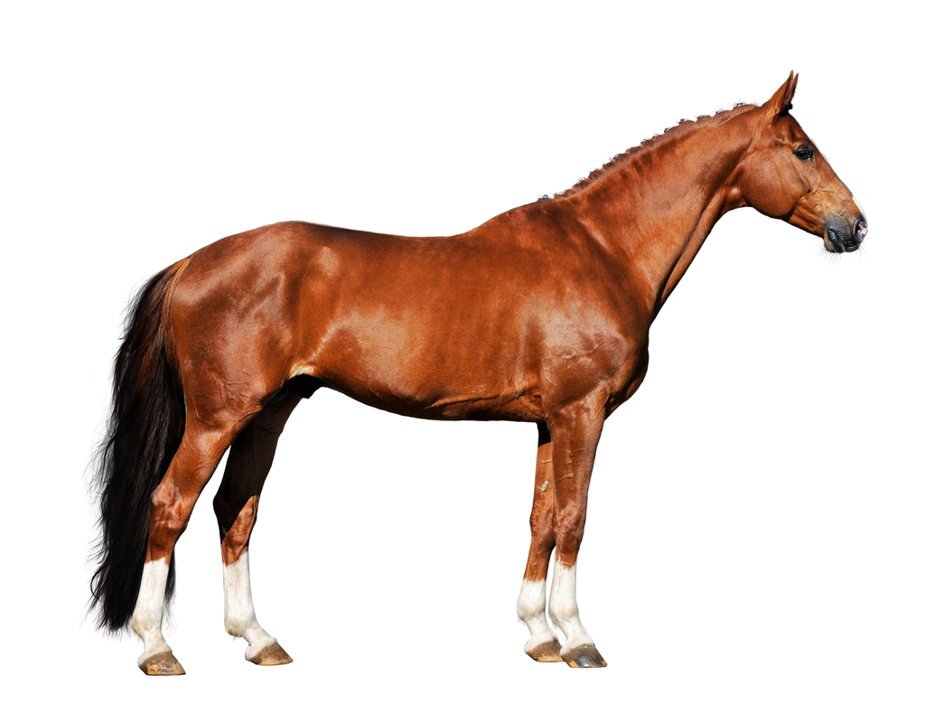
Home » Without Label » Horse Leg Bones Diagram - Forever Horses Anatomy Of The Equine Forleg / The blood supply to and/or from the navicular bone is disrupted.
Horse Leg Bones Diagram - Forever Horses Anatomy Of The Equine Forleg / The blood supply to and/or from the navicular bone is disrupted.
Horse Leg Bones Diagram - Forever Horses Anatomy Of The Equine Forleg / The blood supply to and/or from the navicular bone is disrupted.. Made up of the os coxae, the largest of the flat bones in a horse. Horse stifle joint anatomy via. Horse rear legs the horse leg anatomy in the rear includes the bones of the pelvis (the ilium, ischium and pubic bones), femur, tibia, fibula, metatarsus and the phalanxes. It is made up of the ilium, the ischium, and the pubis.at the junction of these three bones is a cavity called the acetabulum, which acts as the socket of the hip joint.the pelvic cavity is larger in diameter in the mare than in the stallion, providing more room for the foal during birth. The shoulder joint is the articulation between the glenoid cavity of the scapula and the head of the humerus.in the horse, lateral and medial movements of this joint are impossible due to the shape of the humeral head;
This is supposed to demonstrate a change from browsing on bushes to grazing on grass. The wall is made up of the toe (front), quarters (sides) and heel.when the foot is lifted off the ground, the sole and frog are visible, as well as the bars of the wall and the collateral. Get the basics on horse anatomy that every horse owner needs. It is likely that abnormal biomechanical stresses are the basis for the disease. Horse leg structure there are also many fossil remains of horse leg bones.
Disorders Of The Foot In Horses Horse Owners Merck Veterinary Manual from www.merckvetmanual.com The horses legs and hooves are also unique, interesting structures. The eohippus horses part b. A horse's hoof is composed of the wall, sole and frog. Horse rear legs the horse leg anatomy in the rear includes the bones of the pelvis (the ilium, ischium and pubic bones), femur, tibia, fibula, metatarsus and the phalanxes. Movement is therefore limited to flexion and extension. The legs of a horse are made up of a system of muscles, tendons, ligaments, and connective tissue. The arterial supply to the digit and fetlock of the thoracic limb comes mainly from the median palmar artery.the median palmar artery divides in the distal fourth of the metacarpus between thesuperficial and deep digital flexor tendons and the suspensory ligament, to become the medial and lateral digital arteries.part of the deep palmar arch anastamoses with the lateral digital. This system works together to support horses weight when it stands up also works to diminish compression during movement which helps to horse to avoid injury to their limbs.
This is because there are many layers of muscles.
Horse anatomy diagrams legs via. Performance horses tend to suffer from this degenerative disease. Movement is therefore limited to flexion and extension. How many front toes did the oldest horse have? Disorders of the fetlock and pastern include conditions such as fractures, osteoarthritis, osselets, ringbone. The bones of the horse skeleton are held together with ligaments, tendons and muscles. Their leg bones are proportioned differently from those of a human. The bulk of soft tissue is behind the bones. These bones are called the carpal bones, except for the 7th bone which is referred to as the accessory carpal bone. The wall is made up of the toe (front), quarters (sides) and heel.when the foot is lifted off the ground, the sole and frog are visible, as well as the bars of the wall and the collateral. Six small bones make up this joint, and it is often the site of strain and wear and a common location for arthritis. The wall is simply that part of the hoof that is visible when the horse is standing. Images (1) tables (0) videos (0) fetlock is a term used for the joint where the cannon bone, the proximal sesamoid bones, and the first phalanx (long pastern bone) meet.
It also includes the joints of the hip, stifle, hock, fetlock, pastern, and coffin. In this picture it shows the muscles that are closest to the surface of the skin, making them superficial. Diagram of leg bones and joints via. Six small bones make up this joint, and it is often the site of strain and wear and a common location for arthritis. The limbs play a major role in the movement of the horse, with the legs performing the functions of absorbing impact, bearing weight and providing thrust.
Horse Leg Anatomy Form And Function Equimed Horse Health Matters from s.equimed.com Horse body parts diagram, horse skeleton diagram and animal nervous system diagram are some main things we want to present to you based on the gallery title. At this stage of life, even with this exceptionally early development, horses have only 17% of their mature bone mineral content. This is logical, as a horse moves forward the bones of the lower leg shield the soft tissues. How many front toes did the oldest horse have? Get the basics on horse anatomy that every horse owner needs. Which is the oldest horse on the diagram? The power propulsion system and major defensive tool, a horse's rear. It covers the front and sides of the third phalanx, or coffin bone.
Six small bones make up this joint, and it is often the site of strain and wear and a common location for arthritis.
Disorders of the fetlock and pastern include conditions such as fractures, osteoarthritis, osselets, ringbone. It is made up of the ilium, the ischium, and the pubis.at the junction of these three bones is a cavity called the acetabulum, which acts as the socket of the hip joint.the pelvic cavity is larger in diameter in the mare than in the stallion, providing more room for the foal during birth. Their leg bones are proportioned differently from those of a human. The line leading from eohippus to the modern horse exhibits the following evolutionary trends: This is because there are many layers of muscles. Chestnuts horses have small growths on the inside of the front and hind legs called chestnuts. Most bad injuries to lower legs occur when the horse. The small bone that forms the point of the hock is actually similar to the human heel bone. It covers the front and sides of the third phalanx, or coffin bone. Movement is therefore limited to flexion and extension. The bulk of soft tissue is behind the bones. Few animals are as precocious as the horse. Horse body parts diagram, horse skeleton diagram and animal nervous system diagram are some main things we want to present to you based on the gallery title.
The wall is made up of the toe (front), quarters (sides) and heel.when the foot is lifted off the ground, the sole and frog are visible, as well as the bars of the wall and the collateral. How many front toes did the oldest horse have? Develop a better understanding of where leg injuries occur, and the inner workings of the horse hoof. The small bone that forms the point of the hock is actually similar to the human heel bone. The blood supply to and/or from the navicular bone is disrupted.
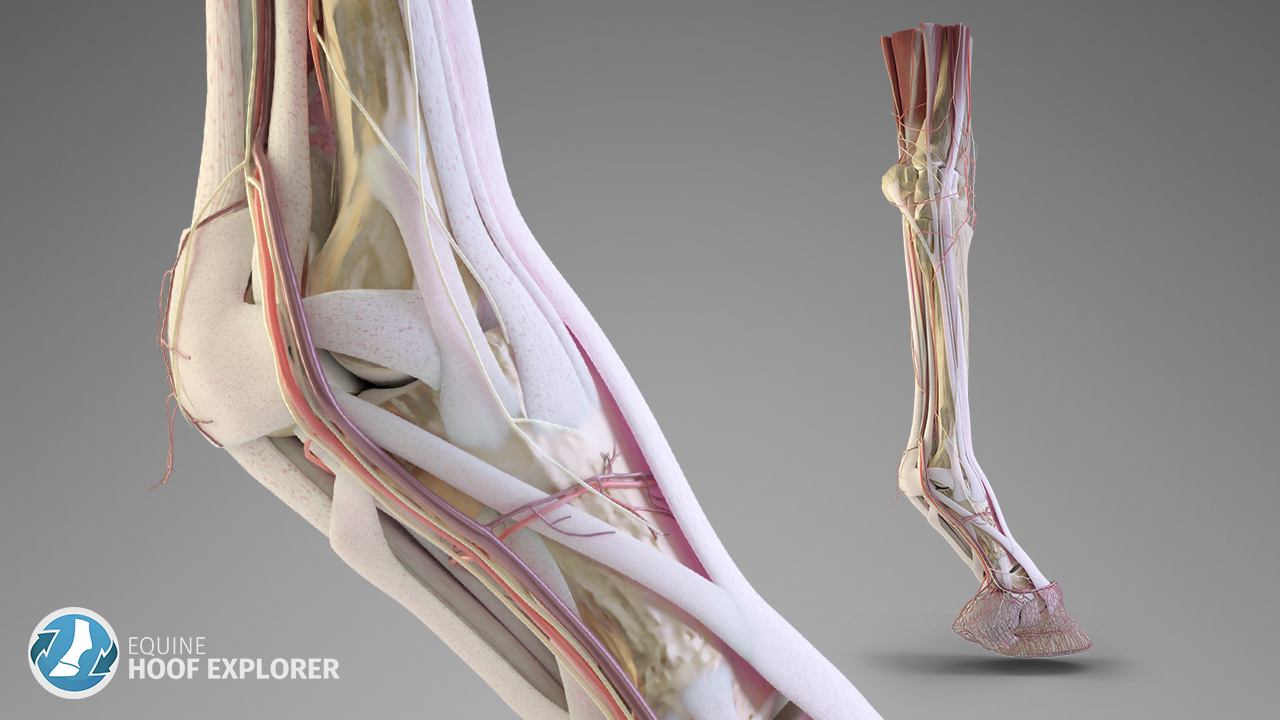
Equine Hoof Explorer 3d Anatomy Software For The Horese Hoof from hoofexplorer.com The ischium forms the point of the buttock. Which is the oldest horse on the diagram? In this picture it shows the muscles that are closest to the surface of the skin, making them superficial. The horse leg anatomy in the rear includes the bones of the pelvis (the ilium, ischium and pubic bones), femur, tibia, fibula, metatarsus and the phalanxes. The legs of a horse are made up of a system of muscles, tendons, ligaments, and connective tissue. The blood supply to and/or from the navicular bone is disrupted. The photograph shows the laminae which keep the hoof wall tightly bonded to the internal structures. Their leg bones are proportioned differently from those of a human.
Human anatomy muscles coloring pages printable via.
The blood supply to and/or from the navicular bone is disrupted. The joint is strengthened by the medial and lateral glenohumeral ligaments. This diagram shows the superficial layer of the tissue. For example, the body part that is called a horses 'knee' is actually the carpal bones that correspond to the human wrist. Their leg bones are proportioned differently from those of a human. It is likely that abnormal biomechanical stresses are the basis for the disease. In the front of the leg, the only thing covering the bones is skin, the extensor tendons (which are very flat) and the suspensory ligaments and fascia. Horse leg structure there are also many fossil remains of horse leg bones. Develop a better understanding of where leg injuries occur, and the inner workings of the horse hoof. The horse leg anatomy in the rear includes the bones of the pelvis (the ilium, ischium and pubic bones), femur, tibia, fibula, metatarsus and the phalanxes. One bone works in relation to another. Performance horses tend to suffer from this degenerative disease. The horses legs and hooves are also unique, interesting structures.
Horse leg structure there are also many fossil remains of horse leg bones leg bones diagram. Inflammation of navicular bone and/or bursa.